
Digital X-ray imaging has made diagnosing orthopedic conditions easier and faster, and the images are much clearer than ever before. We at Skagit Northwest Orthopedics continuously integrate the latest medical and radiography technology to better serve the health of our patients and community.
What is a Digital X-ray?
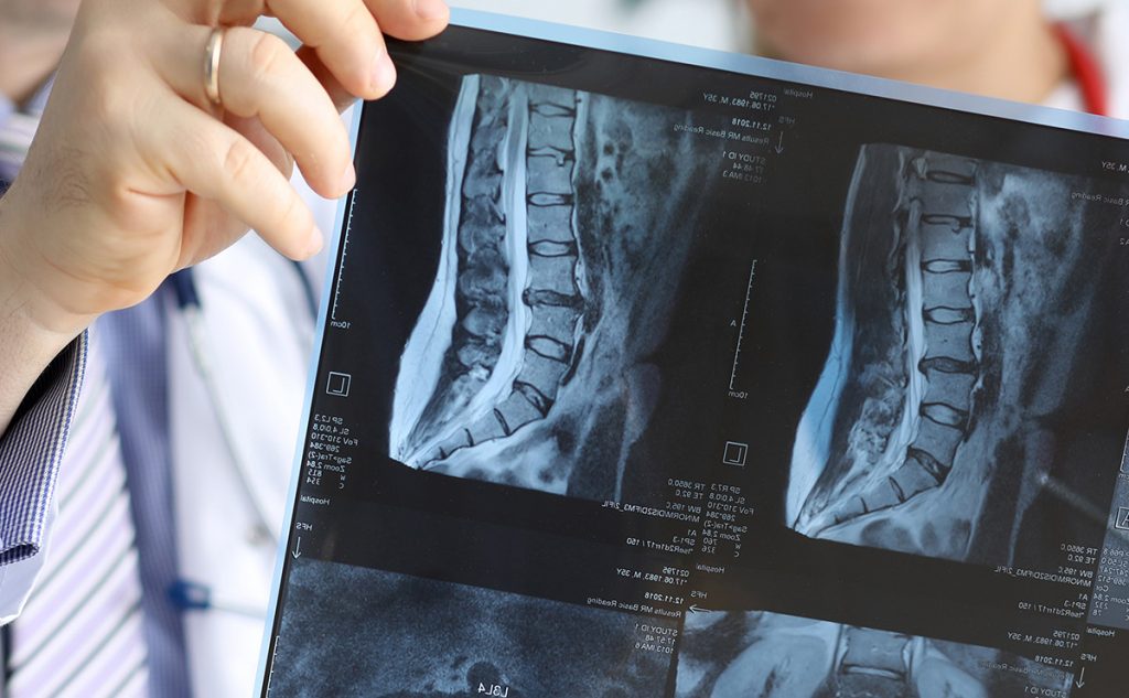
Instead of using film to record the images, like traditional X-rays, digital systems use electronic sensors to capture the X-ray photons that pass through the body. These electronic signals are converted into digital images that are much clearer than the imaging previously used.
Digital X-rays vs. Traditional X-rays
Digital radiography offers several advantages over traditional film-based X-rays, including:
- Faster images: X-ray results can be processed almost instantly, allowing for quicker diagnosis and reduced waiting times.
- Lower radiation exposure: While the amount of radiation used in X-rays is generally low, digital X-ray systems usually require less radiation to produce high-quality images.
- Image enhancement and manipulation: Digital images can be easily enhanced, manipulated, and zoomed in on a computer screen, providing better visualization of specific areas of interest.
- Electronic storage and retrieval: Digital X-ray images can be stored electronically, making it easier to manage and access patient records, and can be easily transferred between healthcare providers if needed.
- Integration with other technologies: Digital X-rays be integrated with other digital medical imaging technologies, such as picture archiving and communication systems (PACS), allowing for seamless sharing and management of medical images.

Why we Use Digital Radiography Imaging
Staying up-to-date with current medical technology serves our patients and allows us to provide greater care. Digital X-rays offer greater insights than traditional ones, but also aid our medical staff with:
- Remote access and consultation: Digital X-rays can be easily transmitted and shared electronically, enabling orthopedic specialists to review images remotely. This is particularly useful for consultations with other specialists or for patients to obtain second opinions.
- Digital measurement and analysis tools: Digital X-ray systems often come with measurement and analysis tools that allow orthopedic doctors to assess angles, distances, and other parameters more accurately, aiding in treatment planning.
What Can It See?
There are many types of imaging technology, each used to see certain types of injuries and aspects of the body. A digital X-ray can show:
- Bone fractures: X-rays are extremely effective in imagining bones, and are commonly used to identify fractures or breaks.
- Joint conditions: They can help diagnose joint-related issues, such as arthritis, by showing changes in the spacing between bones, the presence of bone spurs, or other abnormalities in the joint structure.
- Tumors: Bone tumors and abnormalities in bone structure can be identified.
- Infections: They can be used to detect signs of infection in bones, such as osteomyelitis. Changes in bone density and the presence of soft tissue swelling can be more clearly visible on digital X-ray images.
- Orthopedic conditions: They can be used to assess conditions affecting the musculoskeletal system, such as scoliosis, kyphosis, and other spinal deformities.
- Joint injuries: X-rays are commonly used to evaluate injuries to joints, such as dislocations or misalignments.